Image Challenge #12
01-02-2024
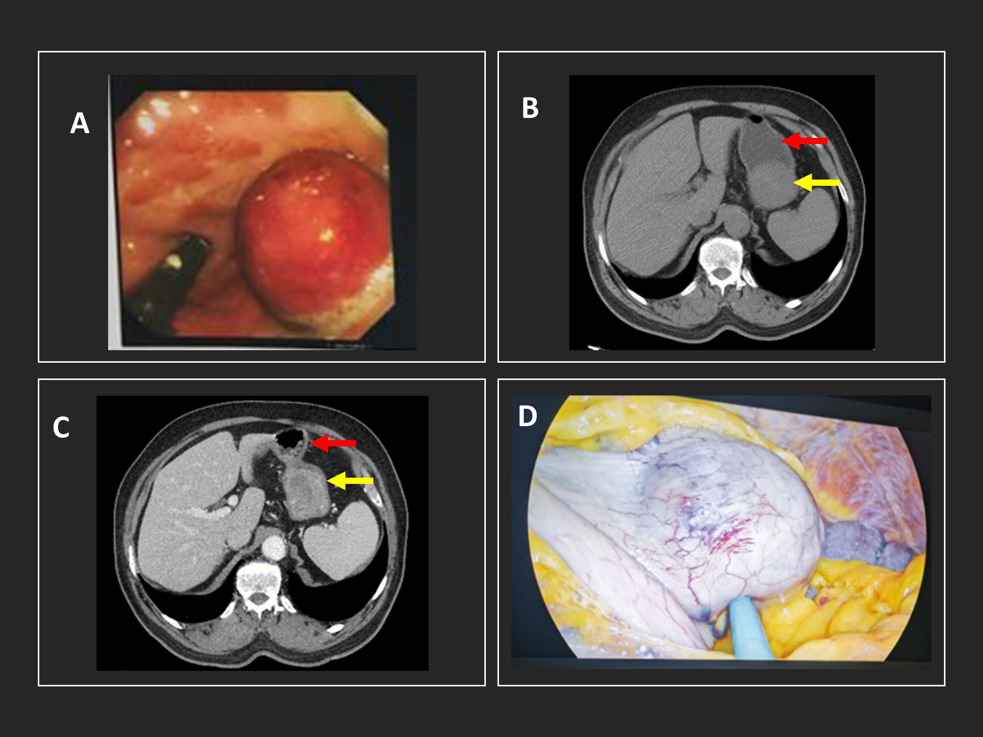
A 76-year-old male presented with melena and anemia. He needed 10 pints of blood. Upper GI endoscopy showed a large fundal submucosal mass projecting into the lumen of stomach (see image A). A CT scan of his Abdomen revealed a well-defined mass arising from gastric fundal wall and avid enhancement with area of necrosis (see image B, C) (red arrow: stomach; yellow arrow: mass). The mass was removed surgically using a laparoscopic technique (see image D).
What is the term used to describe this type of gastric tumor?
Answer: Gastrointestinal stromal tumors (GISTs)
Presented by Dr. Ahmed R. Jawad, Consultant Gastroenterology Surgeon, Hepatology and Gastroenterology Center, Najaf, Iraq.